
My experience at Med Uni Graz was truly incredible – a career highlight. The Department of Macroscopic and Clinical Anatomy team was so welcoming and taught me all about their teaching facilities and specimen collection. I have since left Graz and ventured on to Berlin.
My first medical museum post-Graz has been the Berliner Medizinhistroische Museum der Charité or Berlin Museum of Medical History. Not only do they have an extensive pathology specimen collection, but also a range of historical objects. Understandably, there are photography restrictions in the pathology specimen section, so I’ll only be sharing images of the historical objects.

Context: Museum
I could write at length about the Charité Universitätsmedizin Berlin campus, where the museum is located. It is filled with these eerie-looking red brick buildings, each with its own rich history. For this post, I will focus on the museum. I am taking a tour of the campus on Saturday so watch this space.

The museum’s website has a great section on the history of the building and the collection which I will summarise here. The pathology specimens were collected by Rudolf Virchow, a famous German physician who had a special interest in pathology. He amassed this significant collection not only to assist with medical teaching, but to increase the public’s access to knowledge on health and disease. By 1901, Virchow had collected approximately 23 000 specimens. All he needed now was a place to display them and use them in teaching.
In 1899, the museum opened and under Virchow, had three primary aims:
- Encourage private study of specimens by medical students
- Teach medical students
- Teach the interested public.
Between 1914 and 1939, the museum closed to the public and was used exclusively for medical student teaching. While the collection grew to almost 35 000 specimens, only 1 800 remained at the end of World War II. In addition, the building was severely damaged by bombings. The museum and collection were unused for many decades. Fast forward to the fall of the Berlin Wall. The museum was rebuilt with the aim of becoming a collection for the public. It officially opened in 1998.
Context: Collection

I’ve touched a bit on the pathology collection in the previous section but I wanted to add more context. When you visit the museum today, you can expect to see around 750 pathology and anatomy specimens. The majority are potted specimens but there are a few plastinated also on display.
The specimens are isolated to the second level of the museum, displayed in eight large glass cases. They are divided by body system with a section also on abnormal foetus development. Each system starts with a healthy specimen for context. There is a lot of text accompanying each specimen which is quite overwhelming. If you are interested in reading everything, take your time and have frequent rests to prevent visitor fatigue.
Throughout the rest of the museum are an assortment of medical history objects including Virchow’s work desk, wax moulages displaying a range of pathological conditions, an iron lung, and objects from patients admitted to the hospital.
Context: Display and Layout
In total, there are three levels of exhibitions. The top level displays medical history objects spread across two rooms. One room focuses on the stories of individuals which I will talk about later. The other room traces the history of the hospital and the history of medical inventions such as X-rays and microscopes. Photography is allowed on this level. The next level down is where you will find the pathology and anatomy specimens, moulages, and objects relating to Virchow. The final level is reserved for temporary or travelling exhibitions.

Next to the museum’s entrance are the ruins of Virchow’s lecture theatre. These ruins have been stabilized and the room is now used for presentations, lectures, and events.
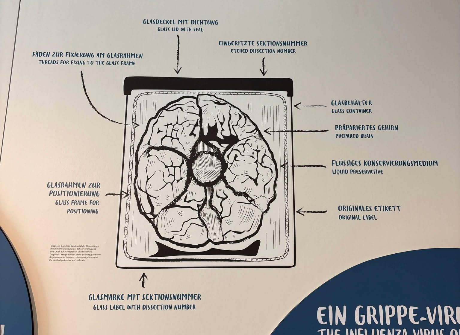
Highlight: Specimen Pot Decal
On the back wall inside the pathology specimen room, there is this incredible decal on the wall. I asked a security guard if I could take a photograph and they very kindly said yes. As you can see, this decal adds so much context to what you are viewing. For someone unfamiliar with potted specimens, I imagine it is a great reference point in understanding the preservation technique.
Highlight: Stories
This one is quite general so I want to provide an example to illustrate. On the top floor of the museum, there is a room displaying patient stories. Running along both sides of the room are five patient stories – so ten in total. Each story is accompanied by a thematic panel, a small display case of personal objects, a large object, and a tall display case with objects to add historical and cultural context,
The example I want to share is the story of polio. The thematic panel tells the story of Hans G., a three-year old patient who suffered from what doctors initially thought was meningitis. As paralysis spread across his body, it became clear that he was suffering from polio. He was transferred to an iron lung for six days. He gradually regained some strength and underwent rehabilitation to re-learn how to walk.
On display next to the panel is Hans’ photo album with images of the hospital, his healthcare team, and his family. There is also a teddy bear that belonged to Hans. The large object in the middle is an iron lung. No matter how many times I see an iron lung, they still shock me. An absolutely enduring symbol of why vaccinations are needed and why they matter. The large display case contains objects relating to the development of a vaccine for polio. There are also some German health posters encouraging people to get the vaccine. The other side of the case has a panel on the disease itself and a leg splint used by a victim of polio for support.


This kind of story telling adds a sense of humanity to the museum. Specimens could have easily been included in these displays but I understand why they were not (photography, etc).
Conclusion
I am looking forward to learning more about the Charité campus on Saturday. When we visited the museum, there was a large school group on a guided tour which interrupted the flow of our visit and made everything quite cramped and crowded. If possible, visit on a weekend. It is a wonderful collection that tells some really significant stories. A definite if you find yourself in Berlin!

Leave a comment